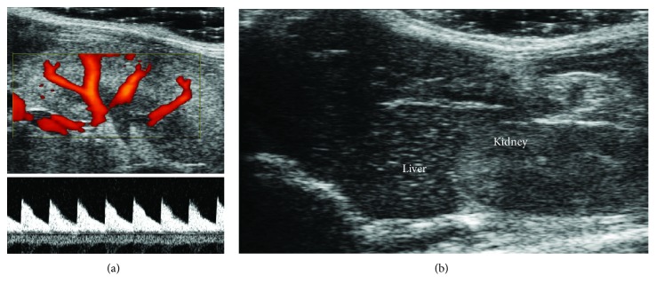
Figure 3.
Kidney and liver ultrasound scan. (a) Power Doppler image was used to localize renal vessels, while PW-Doppler signal was used to measure renal blood flow thus deriving pulsatility and reflectivity indexes. (b) Mean gray levels of the liver parenchyma were compared to mean gray levels of the renal parenchyma.

