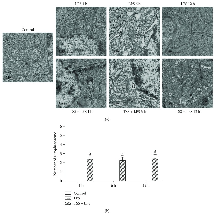
Figure 6.
Autophagosome formation in each group. Representative transmission electron microscope images of each group. (a) A: the control group showed the normal jejunum electron microscopy images; B, C, D: electron microscopy images in jejunum endothelium of LPS-induced mice; E, F, G: the electron microscopy images changed in the jejunum epithelial cells when treated with TSS and LPS. (b) Random cells were chosen to determine the number of autophagosomes/cell for each treatment condition. The black arrowheads indicate membrane-bound vacuoles that are characteristic of autophagosomes. LPS: lipopolysaccharide; TSS: tanshinone IIA sodium sulfonate. Blank column indicates control group; dot column indicates LPS treatment at all observed time points; diagonal column indicates TSS pretreatment at all observed time points; n = 5 in each group. Each bar presents the mean ± SEM. ΔP < 0.05 versus LPS group.

