
Fig 1.
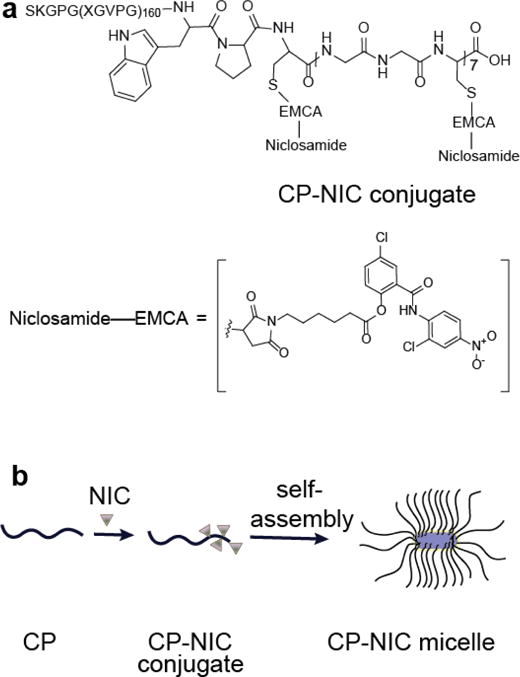
Structure of CP–NIC conjugate and schematic of the structure of CP-NIC nanoparticles. (a) The CP was synthesized by genetically encoded synthesis in E. coli, and conjugated to NIC at the multiple Cys residues at the C-terminal end of the CP. (b) Attachment of the hydrophobic drug NIC (triangles) triggers self-assembly of the CP into cylindrical nanoparticles with a drug-rich (purple) core surrounded by a hydrophilic polypeptide corona (black chains).