FIGURE 6.

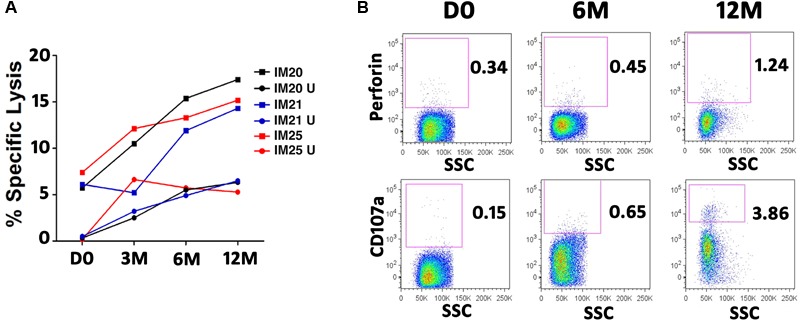
Cytotoxicity of EBNA3-specific T cells against autologous LCLs in three IM patients. T cells were expanded for 5 days in the presence of overlapping peptide pools of EBNA3A or EBNA3B. Autologous B-LCL (target cells) were stained with CFSE and co-cultured with activated purified T cells (effector cells) for 4 h at an Effector/Target ratio of 10:1. Dead cells were discriminated from live cells by propidium iodide (PI). Autologous LCLs were killed by T cells. The percentage of specific lysis is calculated by subtracting the background (LCL co-cultured with unstimulated T cells). (A) Cytotoxicity of EBNA3-specific T cells at different time points in 3 IM patients. PBMC of IM20 and IM21 and IM25 patients were stimulated and expanded with overlapping peptide pools of EBNA3A and EBNA3B, respectively. U, LCL co-cultured with unstimulated T cells. (B) Development of Perforin producing T cells (upper row) and CD107a positive T cells (lower row) in IM25.
