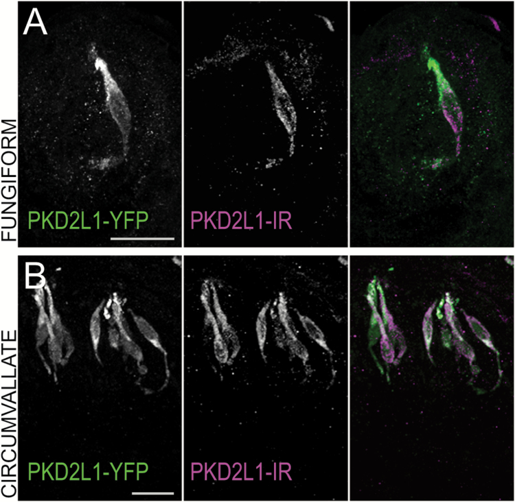
Figure 1.
Transgenic PKD2L1-YFP mice display immunofluorescence in PKD2L1 immunoreactive cells. Confocal z-stack images of (A) fungiform and (B) circumvallate taste buds from a PKD2L1-YFP transgenic mouse showing PKD2L1-YFP fluorescence in green and PKD2L1 immunoreactivity in magenta. Scale bars = 20 µm. In all tissues, the 2 markers are coincident.

