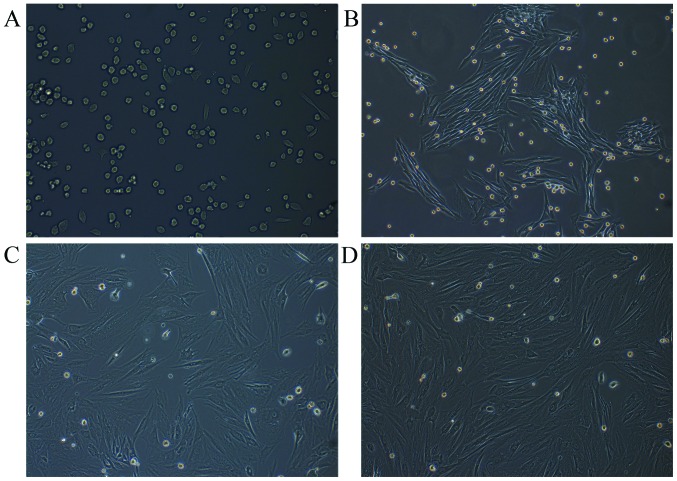
Figure 1.
Morphology of rabbit MSCs. (A) Primary MSCs were cultured for 1 day and circular-shaped cells of uniform size. (B) MSCs at the first passage were cultured for 5 days. (C) MSCs at the second passage were cultured for 5 days. (D) MSCs at the third passage were cultured for 5 days. MSCs were spindle-shaped (fibroblast-like cells) in first, second and third passages after cultured for 5 days. MSCs, mesenchymal stem cells. Magnification, ×100.

