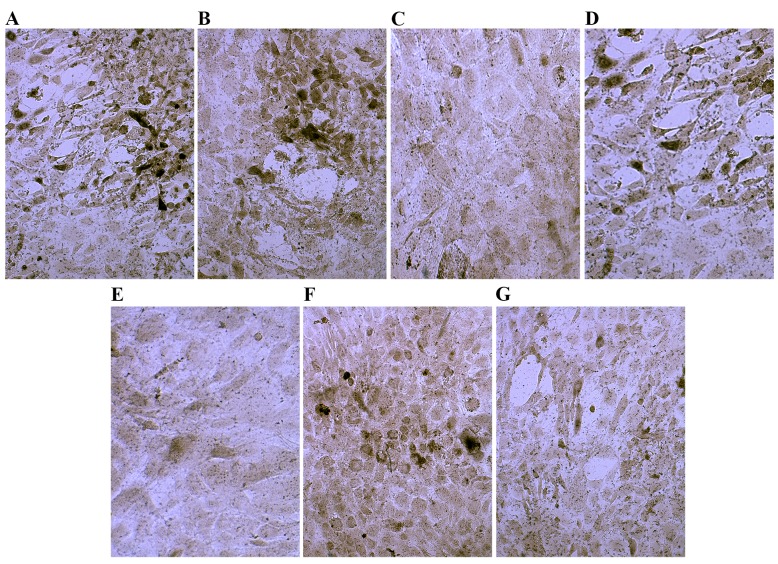
Figure 3.
Phosphatase staining of control cells and cells cultured in osteogenic induction media. Cells cultured in (A) dexamethasone, (B) bone morphogenetic protein 2, (C) 1,25-dihydroxyvitamin D3, (D) transforming growth factor β, (E) platelet lysate, (F) cyclooxygenase 2 and (G) control media. Magnification, ×200.

