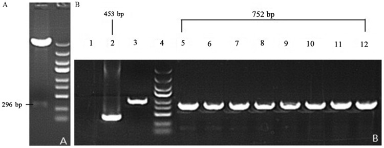
Figure 4.
Gel images of PCR products of recombinant vectors. Following PCR, the double enzyme restriction digestion and agarose gel electrophoresis was performed to determine the fragment size. (A) Enzyme-digested SIRT1-3′-UTR fragments. (B) Lane 1, ddH2O; lane 2, negative control (empty vector self-ligation); lane 3, positive control (GAPDH); lane 4, MW scale (molecular weight of marker protein); lanes 5–12, recombinant SIRT1-3′-UTR luciferase reporter vector plasmid. PCR, polymerase chain reaction; miR-34a, microRNA-34a; SIRT1, sirtuin 1; UTR, untranslated region.

