
Fig. 4. The reduction of CBP stability by FBXL19 impairs histone acetylation and cytokine release.
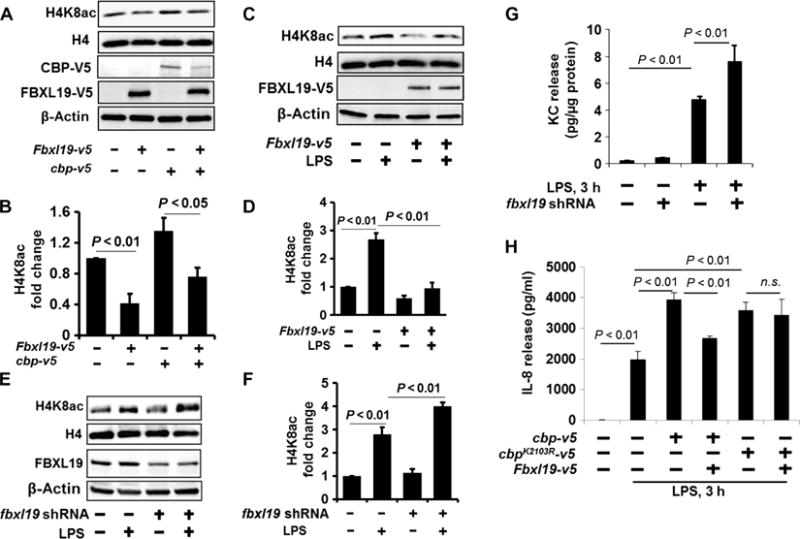
(A and B) MLE12 cells were transfected with cbp-v5 and Fbxl19-v5 plasmids. (A) Forty-eight hours later, isolated histone lysates were analyzed by Western blotting with antibodies against histone H4K8ac and histone H4. Cell lysates were analyzed by Western blotting with antibodies against V5 and β-actin. (B) Determination of relative H4K8ac protein abundance by densitometric analysis of Western blots with ImageJ software. Data are means ± SEM of three independent experiments. P values were calculated by two-way ANOVA and post hoc Tukey’s test. (C and D) HBEpCs were transfected with the Fbxl19-v5 plasmid, and then cells were treated with LPS (10 μg/ml, 3 hours). (C) Isolated histone lysates were analyzed by Western blotting with antibodies against H4K8ac and H4. (D) Determination of relative H4K8ac protein abundance by densitometric analysis of Western blots with ImageJ software. Data are means ± SEM of three independent experiments. P values were calculated by two-way ANOVA and post hoc Tukey’s test. (E and F) MLE12 cells transfected with fbxl19 shRNA (shfbxl19) plasmid. Seventy-two hours later, cells were treated with LPS (10 μg/ml) for 3 hours. (E) Isolated histone lysates were analyzed by Western blotting with antibodies against H4K8ac and H4. (F) Determination of relative H4K8ac protein abundance by densitometric analysis of Western blots with ImageJ software. Data are means ± SEM of three independent experiments. P values were calculated by two-way ANOVA and post hoc Tukey’s test. (G) MLE12 cells transfected with or without the fbxl19 shRNA plasmid. Seventy-two hours later, cells were treated with LPS (10 μg/ml) for 3 hours. KC release was measured by enzyme-linked immunosorbent assay (ELISA). Data are means ± SEM of three independent experiments. P values were calculated by two-way ANOVA and post hoc Tukey’s test. (H) HBEpCs were transfected with the Fbxl19-v5, cbp-v5, or cbpK2103R-V5 plasmid as indicated. Forty-eight hours later, cells were treated with LPS (10 μg/ml, 3 hours). The amount of IL-8 secreted into the medium was measured by ELISA. Data are means ± SEM of three independent experiments. P values were calculated by two-way ANOVA and post hoc Tukey’s test. All Western blots are representative of at least three independent experiments. n.s., not significant.