
Figure 5. In vivo growth of intracellular and total F. tularensis LVS in OS cockroaches.
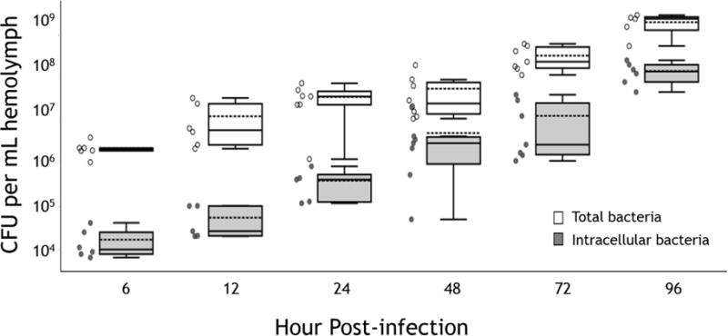
Total (open symbols) and gentamicin-protected (intracellular; shaded symbols) F. tularensis LVS from at least 5 infected OS cockroaches per time point. Bacterial CFU were determined by serial dilution of hemolymph and enumeration on CHOCII agar plates. Intracellular bacterial numbers were determined by injecting gentamicin into infected cockroaches 2 hours prior to each time point. Results from individual insects are shown as open (total CFU) and closed (intracellular CFU) circles. Boxes indicate the median (solid line), mean (dotted line) and interquartile ranges (IQR; box boundaries) for each group. Upper and lower whiskers correspond with the largest and smallest data points, respectively, within 1.5 × IQR for each group.