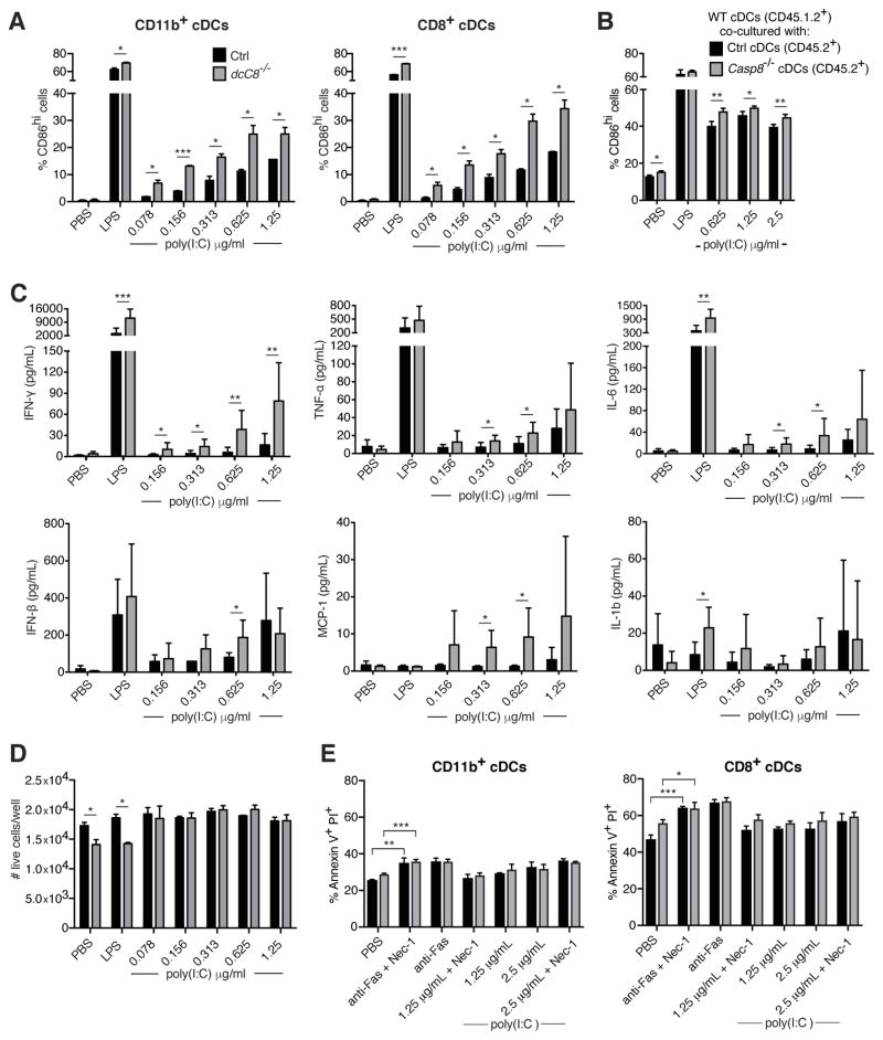
Figure 6.
Caspase-8 deficient cDCs hyperactivate in response to RIG-I stimulation. A) CD11c+ spleen cells were transfected with short-length poly(I:C) for 20 hours, and the expression of CD86 was assessed in CD11b+ and CD8+ cDCs. B) CD45.1.2+ WT CD11c+ cells were co-cultured with CD45.2+ control or Casp8−/− CD11c+ cells in the presence of transfected short-length poly(I:C), and after 16 hours assessed for the expression of CD86. C) CD11c+ cells were isolated from the spleen and transfected with short-length poly(I:C) for 20 hours, and levels of pro-inflammatory cytokines were measured in the cell supernatant by cytometric bead assay. D) The number of live cDCs from A) was assessed by labeling with an amine-reactive dye. E) CD11c+ cells were isolated from the spleen and either stimulated with anti-Fas or transfected with short-length poly(I:C) for 16 hours, with or without Nec-1 pre-treatment. CD11b+ and CD8+ cDCs were then labeled with Annexin V and PI to measure cell death. Data for A, B and D are representative of three independent experiments, with three replicates per genotype. Data for C are representative of two independent experiments, with two replicates per genotype. Data for E are representative of two independent experiments, with three replicates per genotype. Error bars represent S.D.

