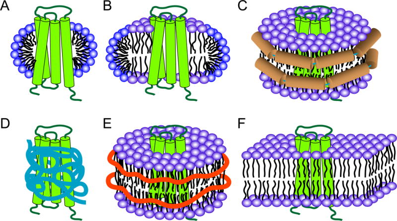
Figure 3.

Membrane mimetics for solution NMR studies of membrane proteins. (A) Micelle, (B) bicelle, (C) nanodisc, (D) amphipol, (E) Lipodisq or SMALP, (F) bilayer in a liposome. A hypothetical four-helical bundle protein is shown in green in all cases. Micelles and bicelles are shown in cut-open views in order to show the differences of these two systems. Membrane scaffold protein (MSP) helices in nanodiscs are shown in brown in (C); amphipols are shown in blue in (D); SMA polymers are shown as orange belts in (E).
