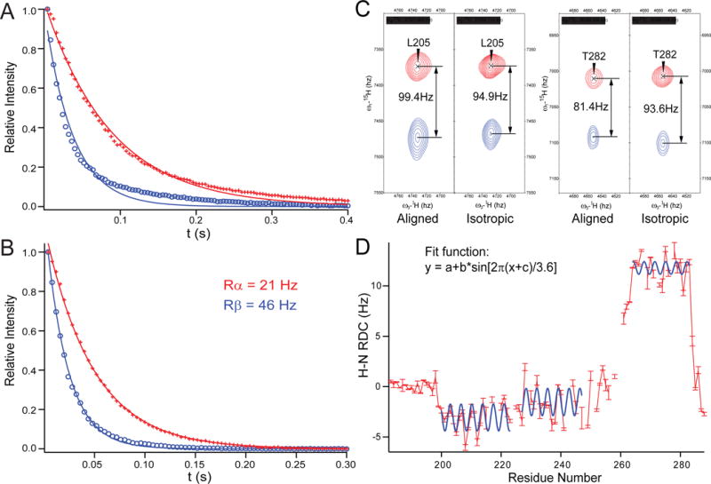
Figure 6.

Syntaxin (183-288) is well structured in DPC micelles. 1D-TRACT NMR experiments of (A) the full-length syntaxin (1-288), and (B) syntaxin (183-288) in DPC micelles. The best-fit single-exponentials to the Rα (red) and Rβ (blue) components are displayed as solid lines. (C) Two pairs of resonances showing different alignments in a 50% negatively charged acrylamide copolymer gel [85] and a final polymer concentration of 4%. (D) Observed H-N RDC values (red bars) versus residue numbers, with three stretches of helical segments (200-224, 227-247, and 264-283) fitted to dipolar waves, i.e., sinusoids of periodicity of ~3.6 residues [86]. Adapted with permission from reference [54].
