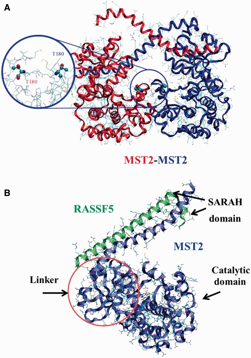
Figure 2.
(A) MST2-MST2 dimer structure (PDB ID: 4LG4). Activation loop and T180 have been highlighted. (B) MTS2-RASSF5 complex from crystal structure (PDB ID: 4LGD) showing the direct interaction between the RASSF5 (green) and the MST2 (blue) SARAH domains. The MST2 kinase domain (blue) is also resolved in the 4LGD crystal structure. Representation of the possible linker between the MST2 kinase and SARAH domains is also shown though, due to its intrinsically disordered nature, it cannot be resolved experimentally. A colour version of this figure is available at BIB online: http://bib.oxfordjournals.org.

