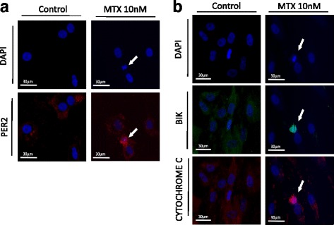
Fig. 5.

PER2, BIK, and CYTOCHROME C expression and morphological changes of the nucleus. a Cells stained with DAPI and anti-PER2 Ab after treatment with MTX (10, 100 nM) or control media for 24 h. Arrowheads indicate apoptotic cells. b Cells stained with DAPI, anti-BIK Ab, and anti-CYTOCHROME C Ab after treatment with MTX (10, 100 nM) or control media for 24 h. Arrowheads indicate apoptotic cells. Bik Bcl-2 interacting killer, DAPI 4′,6-diamidino-2-phenylindole, MTX methotrexate, Per period
