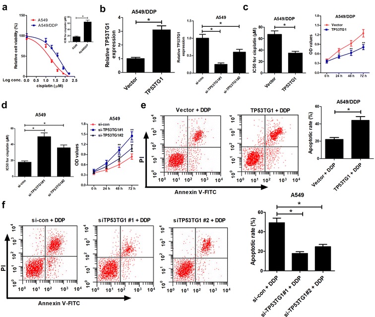
Fig. 2.

TP53TG1 was associated with cisplatin sensitivity in NSCLC cells. a A549 and A549/DDP cells were exposed to different concentrations of cisplatin (1, 10, 20, 40, 80, 160 μM) for 48 h, followed by the determination of cell viability and the calculation of IC50 of cisplatin by MTT assay. b A549/DDP cells were transfected with pcDNA-TP53TG1 and A549 cells were introduced with two individual TP53TG1 siRNAs (si-TP53TG1#1 and si-TP53TG1#2), followed by the detection of TP53TG1 expression by qRT-PCR assay. c pcDNA-TP53TG1-transfected A549/DDP cells were treated with various concentrations of cisplatin for 48 h, and IC50 of cisplatin and cell proliferation capacity were measured by MTT. d si-TP53TG1#1- or si-TP53TG1#2-transfected A549 cells were treated with different doses of cisplatin for 48 h, and IC50 of cisplatin and cell proliferation capacity were monitored by MTT. e Cell apoptosis was evaluated by flow cytometry in pcDNA-TP53TG1-transfected A549/DDP cells after exposed to 60 μM of cisplatin for 48 h. f The apoptotic rate was analyzed by flow cytometry in si-TP53TG1#1- or si-TP53TG1#2-transfected A549 cells after treated with 20 μM of cisplatin for 48 h. Each experiment is repeated three times. *P < 0.05 vs. respective control
