Figure 3.
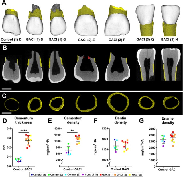
Increased thickness and density of cervical cementum in teeth from generalized arterial calcification of infancy (GACI) subjects. (A) Three-dimensional micro–computed tomography (CT) reconstructions of representative incisors from 1 healthy control and 3 GACI subjects. Cementum layer is highlighted in yellow. Scale bars in panels A to C represent 2.5 mm. (B) Two-dimensional micro-CT cut plane showing hypercementosis in GACI teeth. Cementum layer is highlighted in yellow. Tooth E from GACI subject 2 features calcified pulp stone-like material (labeled in red). (C) Segmented cervical cementum layer used for thickness measurement in control and GACI teeth. Quantitative analyses of control teeth (6 teeth from 4 subjects) and 10 GACI teeth indicate (D) 4-fold significantly increased cementum thickness in GACI vs. control teeth (****adjusted P = 0.00007), (E) 23 % increase in cementum density in GACI vs. control teeth (**adjusted P = 0.009), and no differences in (F) dentin density or (G) enamel density (adjusted P > 0.05 for both). In graphs shown in panels D to G, each individual tooth measurement is displayed with color coding to indicate subject of origin, with mean ± standard deviation shown for 6 teeth from n = 4 controls and 10 teeth from n = 3 GACI subjects. For independent samples t test, multiple teeth from the same individual were first averaged to compare n = 4 control and n = 3 GACI measurements, followed by Benjamini-Hochberg false discovery rate correction for Q = 0.05.

