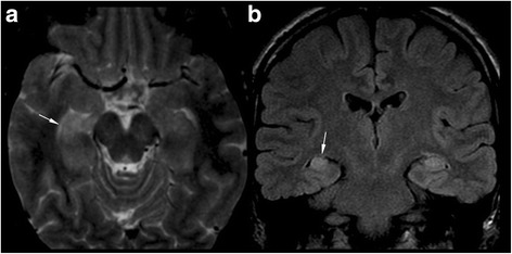
Fig. 3.

Magnetic resonance imaging of patient 1. a An axial FST T2-weighted image and (b) coronal FLAIR image of patient 1 shows slightly asymmetric hyperintense hippocampi (typical UBOs). The right hippocampus shows head volume loss (arrow) and flattened undulations (arrow in a) compared to the left thickened hippocampus. This is consistent with sclerosis
