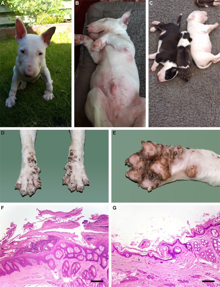
Fig 1. LAD phenotype.
(A) Inflammatory skin lesions in the face of an affected Bull Terrier. (B) Similar lesions in the inguinal region. (C) LAD affected puppy in the middle of two non-affected littermates. A pronounced growth delay and a subtle coat color dilution are visible. (D, E) Fore paws of an LAD affected Bull Terrier puppy at necropsy. Symmetrical scaling and crusting of the skin including interdigital areas and foot pads is visible (F, G) Histopathological micrographs of the junction of interdigital haired skin and digital pad from an affected Bull Terrier puppy (F) and a control dog (G). Marked thickening of the epidermis, excessive layers of non-cornifying epithelium and a large pustule are evident in the affected dog. Hematoxylin-eosin, bar = 400 µm.

