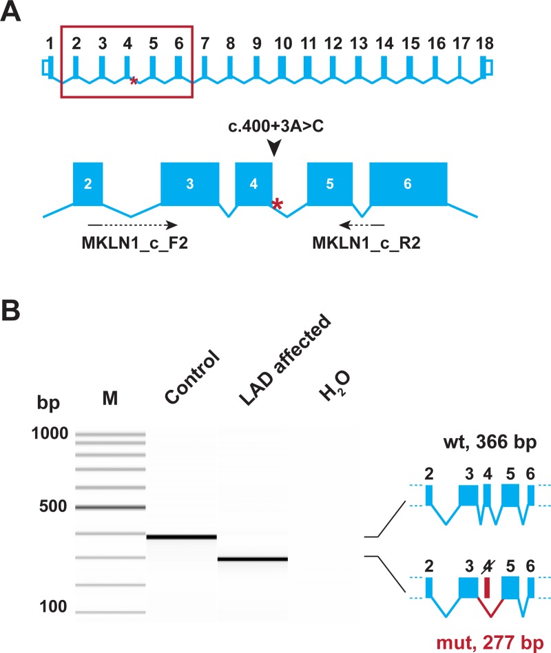
Fig 4. Experimental verification of the MKLN1 splice defect.
(A) The genomic organization of the MKLN1 gene. Exons 2–6 are enlarged and the position of the primers used for RT-PCR is indicated. (B) RT-PCR was performed using skin cDNA from a control and an LAD affected Bull Terrier. The picture shows a Fragment Analyzer gel image of the experiment. In the control animal, only the expected 366 bp product is visible. In the LAD affected dog, a 277 bp product representing a transcript lacking exon 4 is visible. The identity of the bands was verified by Sanger sequencing. Thus, the MKLN1:c.400+3A>C variant leads to complete skipping of exon 4 (MKLN1:r.312_400del89).

