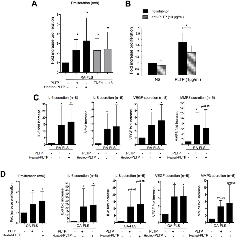
Fig 3. Recombinant PLTP induced FLS proliferation and cytokine production independently of its lipid transfer ability.
(A) RA-FLS were stimulated for 48 hours with 2μg/ml of PLTP or heat inactivated-PLTP (heated-PLTP) and proliferation was evaluated using [3H] thymidine incorporation during the last day of stimulation. Results are expressed as mean fold increase ± SD (n = 8). Statistical differences were assessed by Wilcoxon matched paired test. *p < 0.05 versus unstimulated conditions; TNF-α and IL-1β: positive controls of proliferation. (B) Blockade of PLTP decreased the effect of rhPLTP on FLS proliferation. Cells were treated with PLTP pre-incubated or not with anti-PLTP antibody (n = 5). *p< 0.05 (one-tailed p value), Wilcoxon matched paired test. (C) Effect of PLTP on RA-FLS cytokine production. FLS were stimulated for 24 hours with PLTP or heated-PLTP (2μg/ml). Supernatants were then collected and assessed for cytokines (IL-6, IL-8, VEGF and MMP3) production by ELISA. Results are expressed as mean fold increase ± SD. Statistical differences were assessed by Wilcoxon matched paired test. *p< 0.05 versus unstimulated condition (n = 6 to 8).(D) OA-FLS were treated with either native PLTP or heated-PLTP and analyzed for proliferation (left panel) and cytokine production (right panels) as previously described (n = 5 or 6; *p< 0.05 versus unstimulated condition). RA-FLS and OA-FLS responses were compared using Mann Whitney test and no significant differences could be demonstrated.

