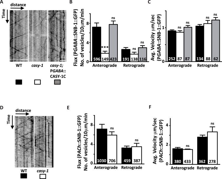
Fig 7. CASY-1 is required for transport of SV precursors in GABAergic motor neurons.
(A) Representative GABAergic::SNB-1::GFP transport kymographs in WT, casy-1 and casy-1; pGABAergic::CASY-1C rescue. For all kymographs, ventral cell body is to the right. Anterograde movement is from right to left. All kymographs are clipped to 10 μm (distance) x 1 min (time) (77x180 pixels). Scale bar, 3 μm. (B-C) Quantification of anterograde and retrograde SV flux in young adult animals. (B) Comparison of mean anterograde and retrograde flux (normalized to a distance of 10 μm and a time of 1 min) between WT, casy-1 mutant and casy-1; pGABAergic::CASY-1C rescue animals. (C) Comparison of mean anterograde and retrograde velocities between WT, casy-1 mutant and casy-1; pGABAergic::CASY-1C rescue animals are shown. casy-1 mutants show significantly reduced anterograde vesicular flux compared to WT animals. Reduced GABAergic anterograde flux was completely rescued by expressing CASY-1C specifically in GABAergic motor neurons. (D) Representative Cholinergic::SNB-1::GFP trafficking kymographs in WT and casy-1 mutant animals. For all kymographs, ventral cell body is to the right. All kymographs are clipped to 10 μm (distance) x 1 min (time). Scale bar, 3 μm. (E-F) Quantification of anterograde and retrograde SV transport in young adult animals (E) Average flux and (F) Average velocity are shown. Flux is defined as the number of moving particles per 10 μm per min. n represents number of particles analyzed for the analysis. Data are represented as mean ± S.E.M. (***p<0.0001 using two-way ANOVA and Bonferroni's Multiple Comparison Test, “ns” indicates not significant in all Figures).

