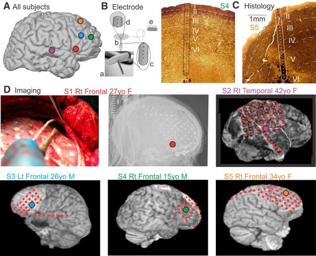
Figure 1.
Electrode localization. A, Approximate locations of laminar probes are mapped on a standard right hemisphere. B, The laminar microarray. Ba, Photograph. Bb, Schematic of the entire array (3600 μm long, 350 μm diameter), and Silastic flap placed on the cortical surface. Bc, Closeup of tip showing the five deepest contacts (each 40 μm diameter, on 150 μm centers). Bd, Closeup of the three most superficial contacts. Be, Connector to amplifiers. C, Histology of the implant sites in 2 subjects, stained with NeuN. D, Imaging of electrode locations, showing placement of the laminar probe under direct visualization in Subject 1 together with postimplant x-rays, and postimplant MRIs in Subjects 2–5. The hemisphere and lobe implanted, age, and gender of the 5 subjects are also shown.

