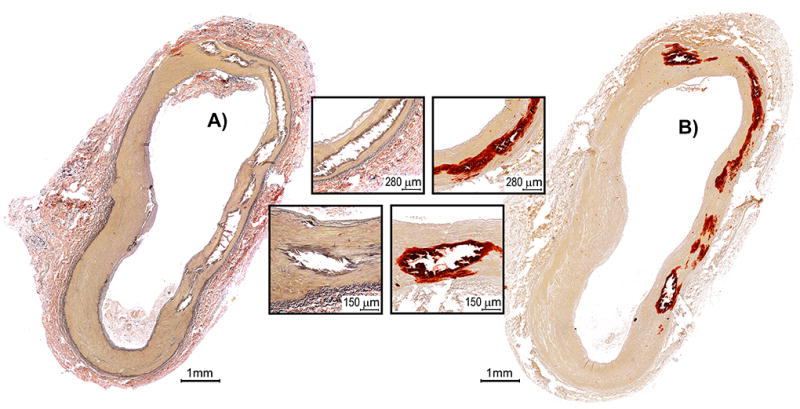
Figure 1.

A) Verhoeff–Van Gieson and B) Alizarin Red S stains of a transverse section of the FPA demonstrating multiple areas of medial calcification (inserts) in the absence of intimal disease. Note that location, amount, and overall calcification burden can be determined using either stain, even though VVG does not specifically strain for calcium and determination relies on tissue artifacts related to calcification. VVG-stained section also provides information on elastic fiber characteristics (strained black).
