Figure 1.
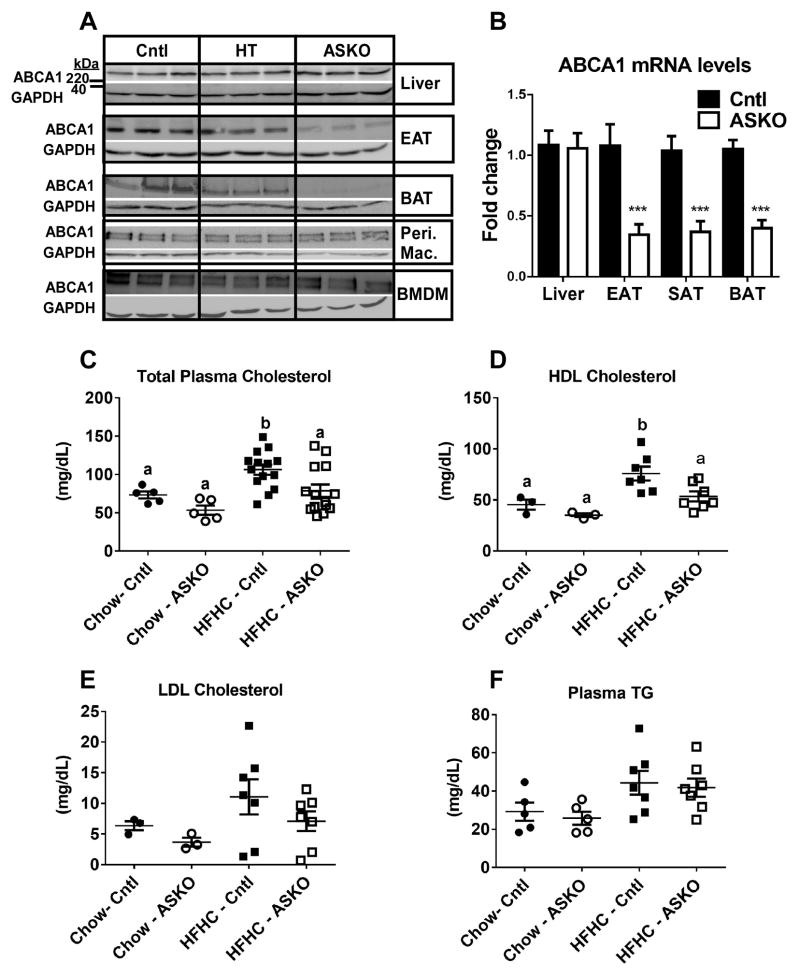
A) Abca1 and GAPDH Western blots of tissues from Cntl, heterozygous ASKO (HT), and ASKO mice. EAT=epididymal adipose tissue, BAT=brown adipose tissue, Peri. Mac.=resident peritoneal macrophages, BMDM=bone marrow-derived macrophages. B) Abca1 gene expression in liver, EAT, SAT and BAT (n=6 per group). *** denotes statistical difference by Student’s t-test, p<0.0001. Plasma concentrations of: C) total cholesterol, D) HDL cholesterol, E) LDL cholesterol, and F) triglyceride (TG) in male Cntl and ASKO mice fed chow for 24 weeks or a HFHC diet for 16 weeks starting at 8 weeks of age. Total plasma cholesterol and TG concentrations were determined from individual plasma samples. LDL and HDL cholesterol concentrations were determined after FPLC fractionation of plasma and enzymatic assay of cholesterol as described in the Methods. Each data point represents an equal volume pooled from 2 mice. Panels C and D, different letters denote statistical difference by ANOVA and Tukey’s multiple comparisons test, p<0.05.

