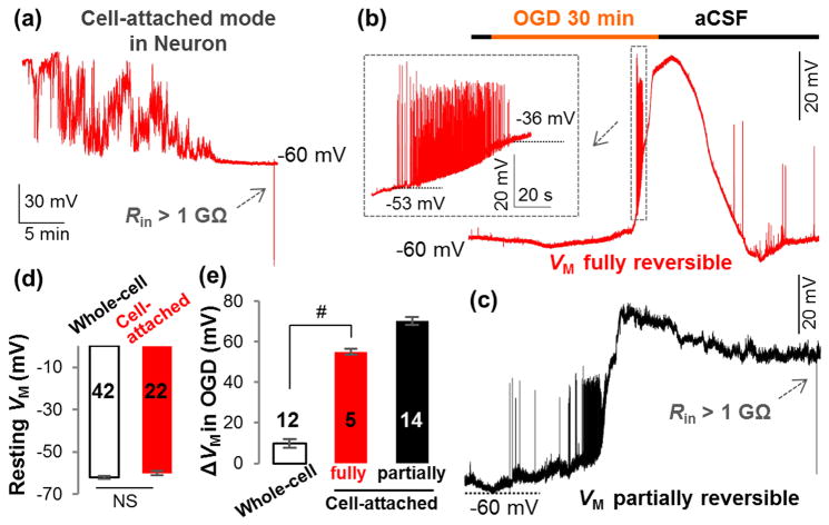
Figure 7. K+ gradient dissipation contributes significantly to OGD-induced neuronal depolarization.
(a) Cell-attached VM recording from a hippocampal pyramidal neuron in situ. Two types of VM responses to OGD are shown in (b) (fully reversible) and (c) (partially reversible). Inset in (b), the neuronal action potentials occurred and vanished during OGD-induced depolarization. (d) The resting VM recorded in cell-attached (a) and conventional whole-cell modes are comparable in pyramidal neurons (P>0.05, t-test). (e) For the neurons with fully reversible VM, OGD-induced depolarization (ΔVM) was significantly greater in cell-attached mode than that of whole-cell mode (P<0.01, t-test). The OGD-induced depolarization from neurons with partially reversible VM is shown in a separate column in (e) but was not included in the statistical comparison.

