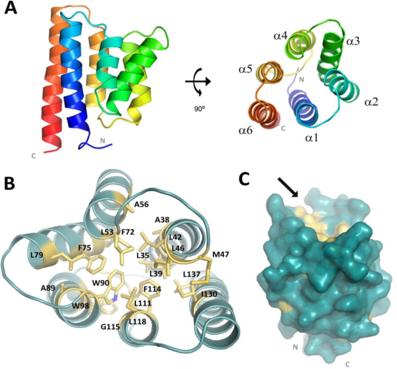
Figure 1.

Overall SpoIIIAB27–153 monomeric structure. (A) Representative SpoIIIAB27-153 structure (cartoon drawing) folds as a six-helices bundle with both N and C termini are in close proximity and facing the same direction (membrane). The molecule is shown in two views, related by a 90° rotation. (B) Conserved residues comprising the SpoIIIAB27-153 hydrophobic core presented as yellow sticks. (C) Surface representation of the monomeric SpoIIIAB27-153 in blue whereas the conserved hydrophobic-core related residues are in yellow. Arrow points toward the small groove located at membrane-opposed protein face and lined with hydrophobic side chains associated with the hydrophobic core.
