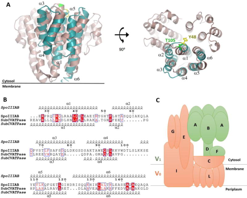
Figure 4.

SpoIIIAB and T. thermophilus V-ATPase shares structural similarity. (A) Structural overlay of SpoIIIAB27-153, in blue, and the T. thermophilus V-ATPase C-subunit, in purple, shown in two views, related by a 90° rotation. SpoIIIAB overlaps to the first domain of the V-ATPase (residues 78-183). Specific and functionally conserved residues that are facing the cytosol are presented as sticks, C-subunit T105 in green and SpoIIIAB Y48 in yellow and the numbers of SpoIIIAB helices are numbered. (B) Sequence alignment of SpoIIIAB27-153 and the V-ATPase C-subunit, residues are numbered according to SpoIIIAB. Top and bottom secondary structures are presented according to their crystal structures. (C) Schematic representation of the T. thermophilus V-ATPase subunits composition. The V1 and V0 domains are colored in green and orange, respectively.
