
Figure 2. Activation of PI3K signaling following sciatic nerve crush.
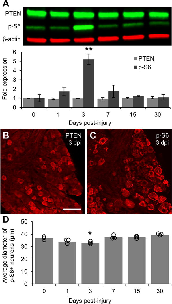
A: Representative Western blot images of PTEN and p-S6 staining from L4/5 DRG lysates with graphical representation of data normalized to 0 day uninjured controls (n = 3 DRG pools per time point). β-actin was used as a loading control. PI3K signaling is greatly elevated at 3 days post-injury as shown by a five-fold increase in the level of p-S6 as compared to controls. p-S6 levels in DRG lysates returned to baseline within 7 days of injury. B: Representative image of PTEN expression in DRG 3 days post-injury showing that injury does not alter the localization of PTEN in small diameter neurons. C: Representative image of p-S6 in DRG 3 days post-injury showing sustained phosphorylation of S6 in large diameter neurons and increased S6 phosphorylation in smaller diameter neurons. D: Graphical representation of the average diameter of neurons positively stained for p-S6 (n = 3 per time point). Individual data points are indicated by hollow circles. ** p < 0.01 vs 0 day uninjured controls in A. * p < 0.05 vs 0 day uninjured controls and 7, 15, and 30 days post-injury in B. Scale bar = 100 μm.