
Figure 2.
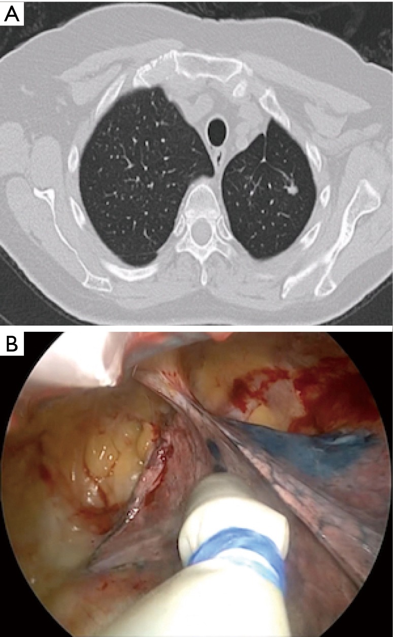
Patient 10. (A) CT scan of a lady with previous history of left upper lobectomy for lung cancer and presented 3 years afterward with an 8-mm pulmonary nodule in the remaining lobe; (B) the Geiger counter probe examines the lung surface, where also methylene blue is visible, during left uniportal VATS radioactivity guided wedge resection. VATS, video-assisted thoracoscopic surgery.