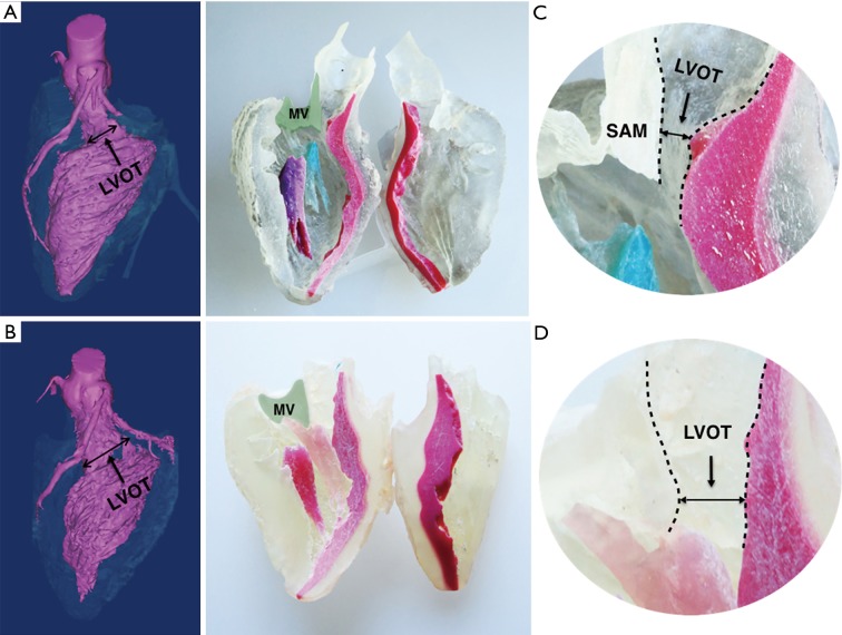
Figure 2.
3D model of hypertrophic obstructive cardiomyopathy in longitudinal section. 3D Model of patient before (above) and after (below) myectomy procedure. (A) The 3D virtual model shows LVOT was significantly enlarged after the procedure; (B) the IVS was colored red and the green shaded area marks the mitral valve area in the corresponding 3D-printed model. The solid line indicates the potential incision of myectomy and the hypertrophic IVS which disappear postoperatively; (C) the narrowed LVOT (the channel between the dashed lines) and systolic anterior motion of mitral valve was illustrated in the larger version of the LVOT; (D) the enlarged LOVT after the myectomy. LVOT, left ventricular outlet tract; IVS, interventricular septum; MV, mitral valve; MVA, anterior leaflet of mitral valve.

