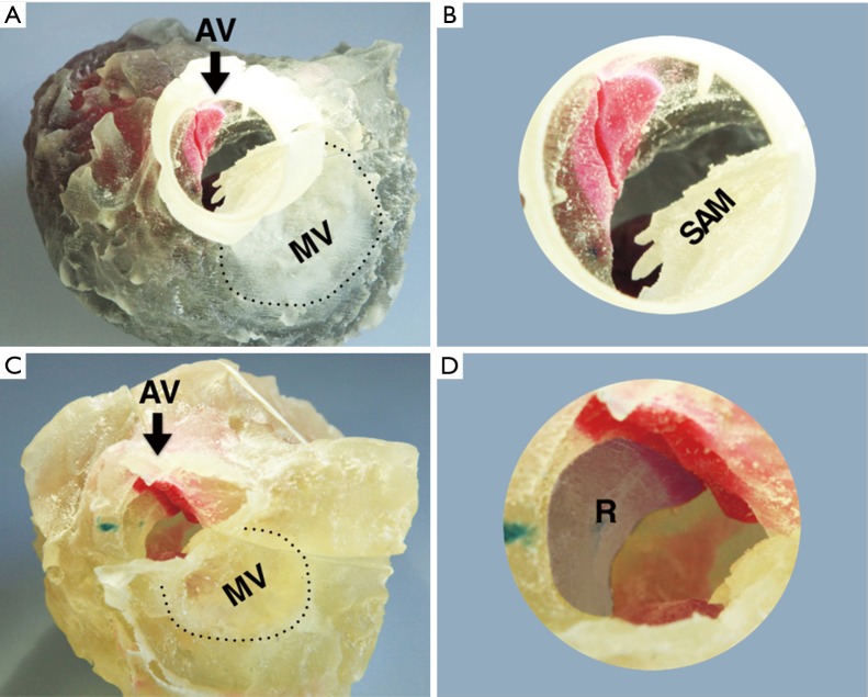
Figure 3.
The superior aspect of the 3D model of hypertrophic obstructive cardiomyopathy. 3D Model of patient before (A,B) and after (C,D) myectomy procedure. The images show the superior aspect of the left heart which the aortic valve is removed. The dashed line indicates the mitral valve annulus. The anterior leaflet forward motion toward the IVS is also obvious in the larger version of the aortic root. The shadow area marked R indicated the removed hypertrophic IVS in the operation. AV, aortic valve; MV, mitral valve; SAM, systolic anterior motion; IVS, interventricular septum; R, resect part of hypertrophic IVS.

