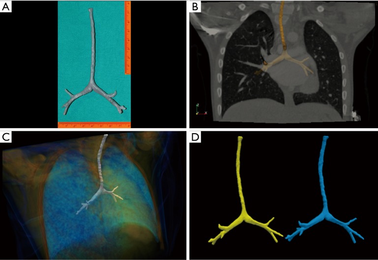
Figure 3.
Preoperative reconstruction. (A) Three-dimensional printed trachea model 1:1 size; (B) fusion image between STL model obtained from the segmentation of the patient’s tracheal images from the CT scan and sagittal MPR projection of DICOM CT volume; (C) fusion image between STL model and volume rendering of CT volume; (D) right side (blue image): STL model of the patient’s tracheal images from the CT scan. Left side (yellow image): STL model obtained from segmentation of the printed model CT scan. To note the perfect matching between the geometry that helps to validate the dimensions of the printed model. STL, stereo lithography; CT, computed tomography; MPR, multi planar reconstruction.

