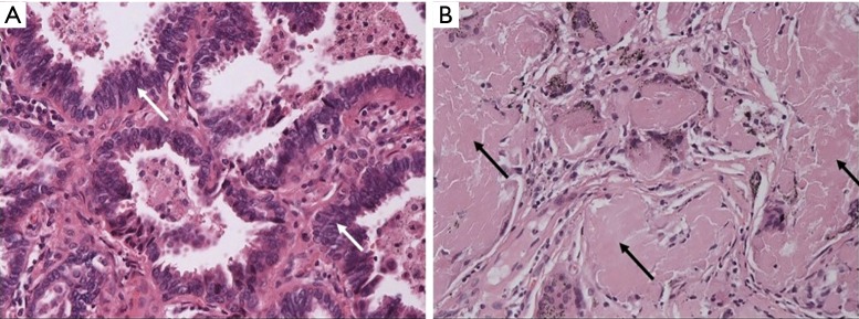
Figure 2.
Pathological findings. (A) The lung adenocarcinoma (white arrows, hematoxylin-eosin staining, 200×); (B) amyloid deposits (black arrows) within the subpleural cyst wall with occasional foreign body giant cells and mild infiltration of chronic inflammatory cells (hematoxylin-eosin staining, 200×).

