
Figure 2.
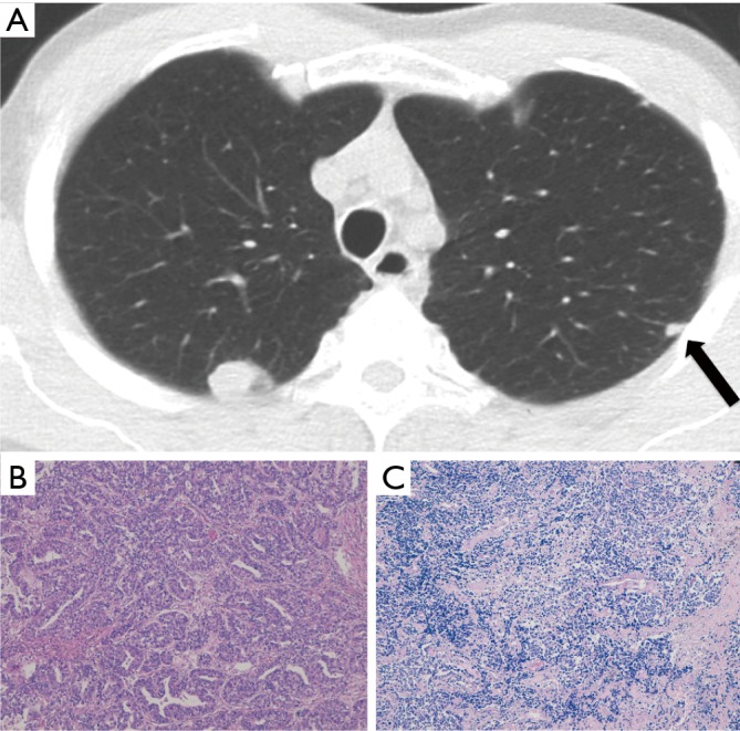
Case II (Table 1). Adenocarcinoma and small cell lung cancer in a 61-year-old smoker both detected at baseline LDCT screening round. They appeared as a rounded lung nodule in the RUL and a subpleural small solid nodule (arrow) in the LUL (A). Haematoxylin and eosin histologic staining demonstrate invasive adenocarcinoma, acinar predominant (original magnification ×200) (B) in the RUL lesion and small cell lung carcinoma (original magnification ×100) (C) in the LUL lesion.