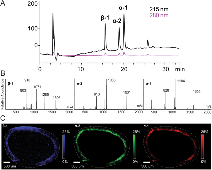
Figure 2.
The discovery of nemertides. (A) RP-HPLC-UV trace of a high molecular weight fraction after SEC, showing nemertides α-1, α-2 and β-1. (B) MS spectra of the three peptides β-1 (left), α-2 (center) and α-1 (right). (C) The spatial distribution of nemertide β-1 (blue, 6419.821 ± 0.5 Da), α-2 (green, 3260.738 ± 0.5 Da) and α-1 (red, 3308.767 ± 0.5 Da) by MALDI-MSI (15 µm resolution) of a transversal section from the mid region of L. longissimus.

