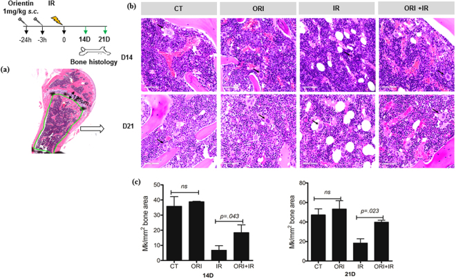
Figure 3.
The bone histology shows an increased megakaryocyte number. MK scoring was done by a subjective analysis of three adjacent high power (60x) microscopic fields for each H&E stained bone section. (a) Megakaryocytes/2 mm2 tissue area was scored 125 µm away from the growth plate for a total area of 2 mm2. (b) Representative tissue sections depict effect of Orientin on megakaryocytes on day 21 post 6 Gy TBI in mice (C57BL/6 8–10 week old). Black arrow denotes MK cells. Scale bar 50 µm (c) MK count from two independent experiments scored after indicated days of 6 Gy exposure (n = 5 each group).

