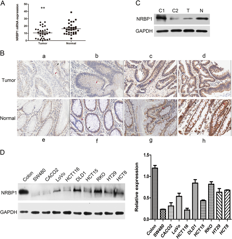
Fig. 1. Expression of NRBP1 is downregulated in CRC.
a qRT-PCR analysis of the expression levels of NRBP1 in 30 pairs of fresh-frozen primary CRC tissues and their matched normal tissues adjacent to cancer tissues. Three experiments were done. NRBP1 mRNA expression was normalised to β-actin mRNA, which used as the internal reference. The data are expressed as the mean ± standard deviation. (**P < 0.01). b IHC staining to detect NRBP1. a Weak staining of NRBP1 in CRC tissues. b–d More intense staining of NRBP1 in CRC tissues. e Weak staining of NRBP1 in normal colorectal tissues. f–h More intense staining of NRBP1 in normal colorectal tissues. Red arrow indicates cytoplasm staining. Black arrow indicates nucleus staining. c The expression levels of NRBP1 protein were analysed by western blot in CRC tissue and matched normal colorectal tissue. GAPDH was used as loading control. N normal tissue, T tumour tissue, C1 protein from stomach tissue was used as positive control, C2 protein from thymus tissue was used as negative control. d Western blot was used to investigate NRBP1 protein expression in nine CRC cell lines and normal colorectal tissue

