Figure 3.
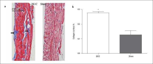
Left superior pulmonary vein (LSPV) fibrosis in 2K1C hypertensive and sham-operated rats (a) Representative examples of Masson’s trichrome-stained LSPV sections from 2K1C hypertensive and sham-operated rats (original magnification 200×, arrows indicate areas of fibrosis). (b) Percentage fibrosis measured as the area that was stained blue as a percentage of the total area using Image-Pro Plus 6.0
Data are expressed as mean±SD. *p=0.016 vs. sham-operated group (n=6)

