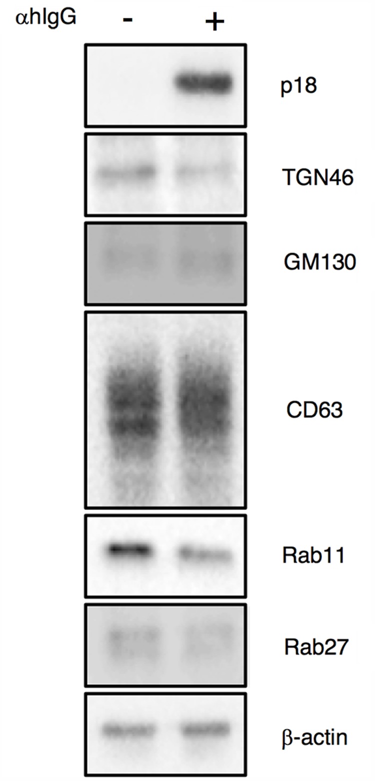
FIGURE 7.

Expression of organelle markers in Akata+ cells treated with αhIgG. Akata+ cells were treated with or without αhIgG for 16 h. Total cell lysates were analyzed by Western blotting with antibodies against p18, TGN46, GM130, CD63, Rab11, Rab27a, or β-actin.
