
FIG 3.
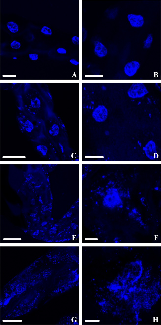
DAPI-stained nuclear DNA of D. citri midguts viewed using confocal microscopy. (A and B) Class 0 (normal) nuclear DNA phenotypes are often solid blue circles and are distinguished by a cohesive edge with semicircular shape. (C and D) Class 1 nuclear DNA phenotypes are irregularly shaped, with the edges tending to show mild breakage or splintering. (E and F) Class 2 nuclear DNA phenotypes still show a noticeable density of DNA reminiscent of the circular phenotype from class 0 but are fragmenting from all points and beginning to disperse. (G and H) Class 3 nuclear DNA phenotypes are highly fragmented with no intact cohesion remaining to suggest that there was a nucleus, as the DNA appears to have spread throughout the cytoplasm. Scale bars: 25 μm (A, B, and G), 50 μm (C), 75 μm (D and E), and 10 μm (F and H).