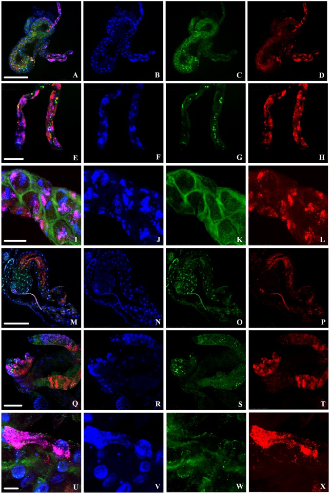
FIG 8.
Localization of “Ca. Liberibacter asiaticus” (green) and Wolbachia (red) in infected D. citri gut cells with DAPI staining of nuclei (blue). Infected adult guts (A to L) and infected nymph guts (M to X) were stained using FISH. (A, E, I, M, Q, and U) Overlays of “Ca. Liberibacter asiaticus,” Wolbachia, and nuclei. Rows 1 to 3 (from the top) are progressively enlarged images of adult gut cells, and rows 4 to 6 are progressively enlarged images of nymph gut cells. Scale bars: 250 μm (A to D and M to P), 75 μm (E to H and Q to T), 50 μm (I to L), and 25 μm (U to X).

