FIG 3.
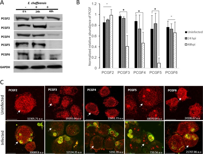
PCGFs redistribute to the ehrlichial vacuole during infection and result in a decreased total cellular level of PCGF isoforms. (A) Western blot analysis of THP-1 (E. chaffeensis-infected and uninfected) whole-cell lysates (0, 24, and 48 hpi) using isoform-specific anti-PCGF antibodies. Whole-cell lysates were subjected to SDS-PAGE, and the amount of each PCGF isoform was detected using chemiluminescence; GAPDH was used as a loading control. (B) GAPDH-normalized relative abundance of PCGF isoforms in uninfected and E. chaffeensis-infected THP-1 cells at 24 and 48 hpi. Error bars indicate standard deviations between experiments (n = 3; Student's two-tailed t test; *, P ≤ 0.05). (C) Confocal laser micrographs showing decrease in PCGF isoforms (red fluorescence) in E. chaffeensis-infected cells at 48 hpi (bottom panel) compared to those in uninfected cells (top panel); scale bar, 10 μm. Arrows indicate specific cells for which the total cell fluorescence was calculated and which is shown on the bottom right corner of the image.

