Figure 3.
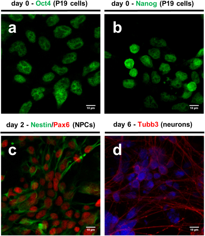
In vitro differentiation of P19 cells mimics early processes in mammalian neuroectodermal development in vivo. (a) and (b) Confocal images of P19 cells stained with anti-Oct4 and anti-NANOG antibody (FITC-green), respectively. (c) Confocal images of P19-derived neural progenitor cells (NPCs) on the second day of neuronal induction upon plating on adhesive tissue culture dishes stained with anti-Pax6 (FITC-green) and anti-nestin (FITC-Texas). (d) Neuronal precursors become post-mitotic and undergo end-terminal differentiation, observed by confocal microscopy of neurons stained with anti-Tubb3 on day 6 of neuronal differentiation upon plating on adhesive tissue culture dishes. Cell nuclei were counterstained with DAPI. Images of immuno-staining and DAPI staining were overlaid. Scale bar represents 10 micrometers.

