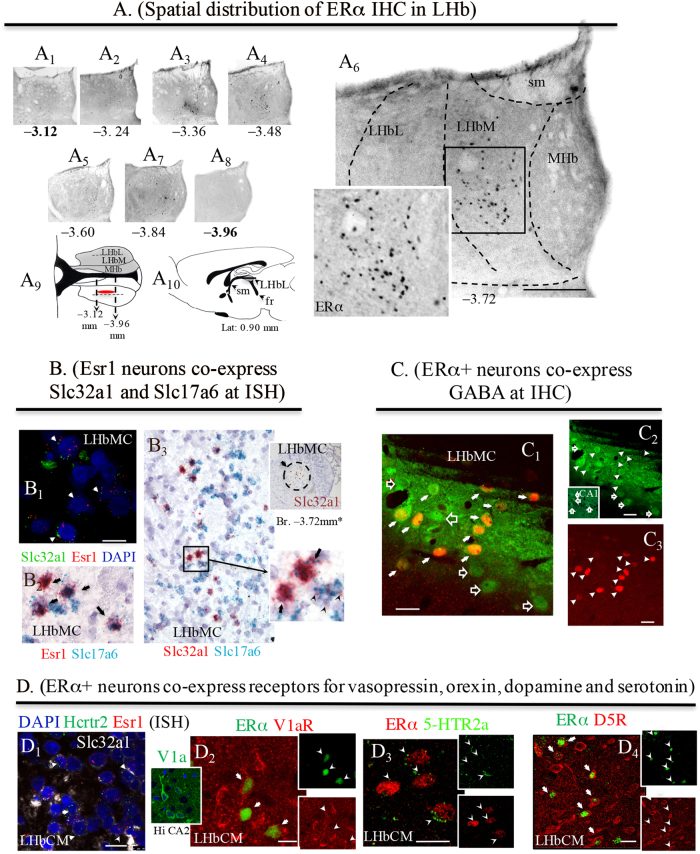
Fig. 2. GABAergic estrogen-receptive neurons (GERNs) localization, mRNA expression by ISH and somatic receptor expression by IHC.
a Serial coronal sections showing the ERα immunolabelling at the Bregma rostro-caudal coordinates (numbers in mm under the photomicrographs). Boxed area in A6 at higher magnification showing the exclusive nuclear labeling pattern. The bold numbered levels (A1 and A8), were chosen to show that no positive labeling was found in either rostral or caudal directions. A9: a horizontal view of the distribution of estrogen-receptive cells was symbolized by the red oval. A10: sagittal view of rat brain atlas, modified from Paxinos & Watson86, at lat. 0.90 mm, where lateral habenula (LHb) is symbolized with a gray shade and A9 plane was symbolized with a horizontal line. b In situ hybridization using multiple RNAscope methods. B1: multiplex fluorescence method, Esr1, gene that encodes ERα (red punctuated labeling) co-expressed with Slc32a1, gene that encodes VGAT (green punctuated labeling); arrows indicate the double-labeled cells; B2: duplex method, Esr1 (red punctuated labeling) co-localization with Slc17a6, gene that encodes VGLUT2 (green punctuated labeling); arrows indicate the double-labeled cells; B3: with duplex method, Slc32a1 encoding VGAT (red punctuated labeling) shows complete co-localization with Slc17a6 encoding VGLUT2 (green punctuated labeling); arrows indicate the double-labeled cells. Inset of B3, Slc32a1 expression in a sexually active (SA) rat LHb, Br. −3.72 mm (brown labeling, single chromogenic-Brown method-RNAscope). *Note the similarity with ERα expression in A6. c Indirect immunohistochemistry showing the GABAergic nature of the ERα+ neurons (red) in a SA rat brain. The GABA antibody (green, Sigma, A0310) produced characteristic surface labeling (C1, C2: the strip-like image was produced by Vibratome slicing leaving the brain section with an uneven surface). d ERα+ neurons co-express receptor/receptor subtypes for vasopressin, orexin, dopamine, and serotonin. D1: In situ hybridization using RNAscope-multiplex method targeting Esr1 (red dots), Slc32a1 (white dots) and Hcrtr2, gene encodes the orexin receptor 2 (green dots). D2, D3: Indirect immunofluorescence reactions, showing that the ERα-IR cells co-expressed vasopressin receptor V1a (inset showing the V1a antibody labeling pattern in temporal hippocampus CA2 cells body layer. See also SI Fig. 6 for more information about this antibody), dopamine receptor D5R (also called D1Rb) and serotonin receptor 5-HTr2a, respectively. Scale bars: A5 and b: 500 µm and rest: 10 µm

