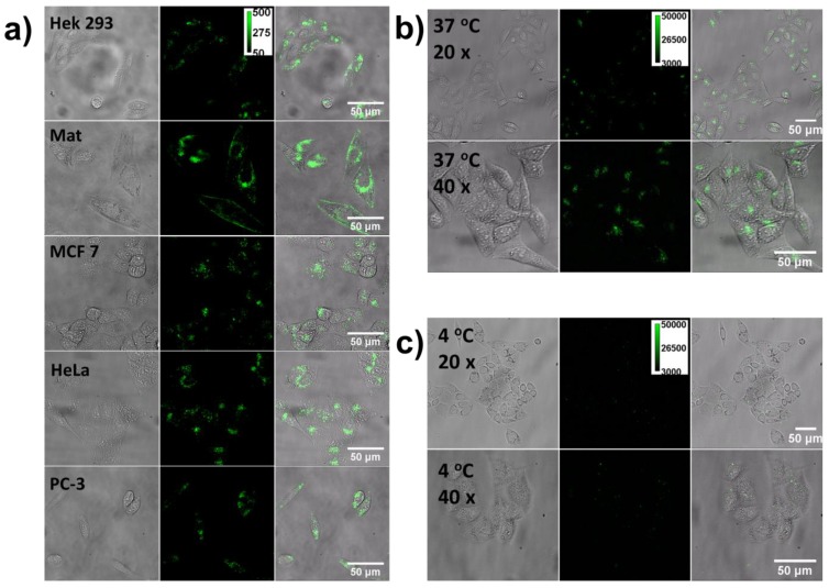
Figure 4.
The cellular uptake efficiency assay. a) Five kinds of cells were co-cultured with the CPNPs for 2 h and then imaged with a fluorescence microscope. Pictures from left to right are the bright-field, fluorescence and merged microscopic images respectively. b) and c) are the fluorescence images of the cells co-cultured with CPNPs for 2 h at 37 and 4 oC respectively.

