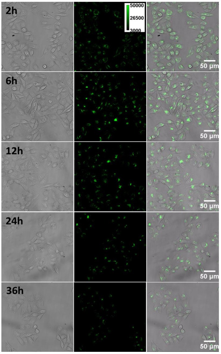
Figure 6.
The tempo-spatially resolved microscopic images of CPNPs inside the cell. Pictures from left to right are the bright-field, fluorescence and merged microscopic images of the cells. After internalized by the cell, CPNPs inside the cytosol could gradually assemble close to the periphery of the nucleus as time goes on.

