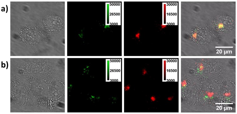
Figure 7.
Lysosome staining experiments. a) The cells co-cultured with the CPNPs for 4 h and then stained with lyso-Tracker Red. Pictures from left to right are the bright-field, green channel fluorescence (from CPNPs), red channel fluorescence (from the lysosome tracker) and merged images of living cells. As noted, the majority of the fluorescent spots from green and red channels are overlapped well, demonstrating that most of the CPNPs are inside the vesicles after internalized by the cell. b) The same staining experiment after co-incubating the cell with CPNPs for 12 h. As demonstrated in the merged image, the fluorescence from the green and red channels cannot be well merged together. However, those signals from the CPNPs are still staying close to the periphery of the nuclei.

