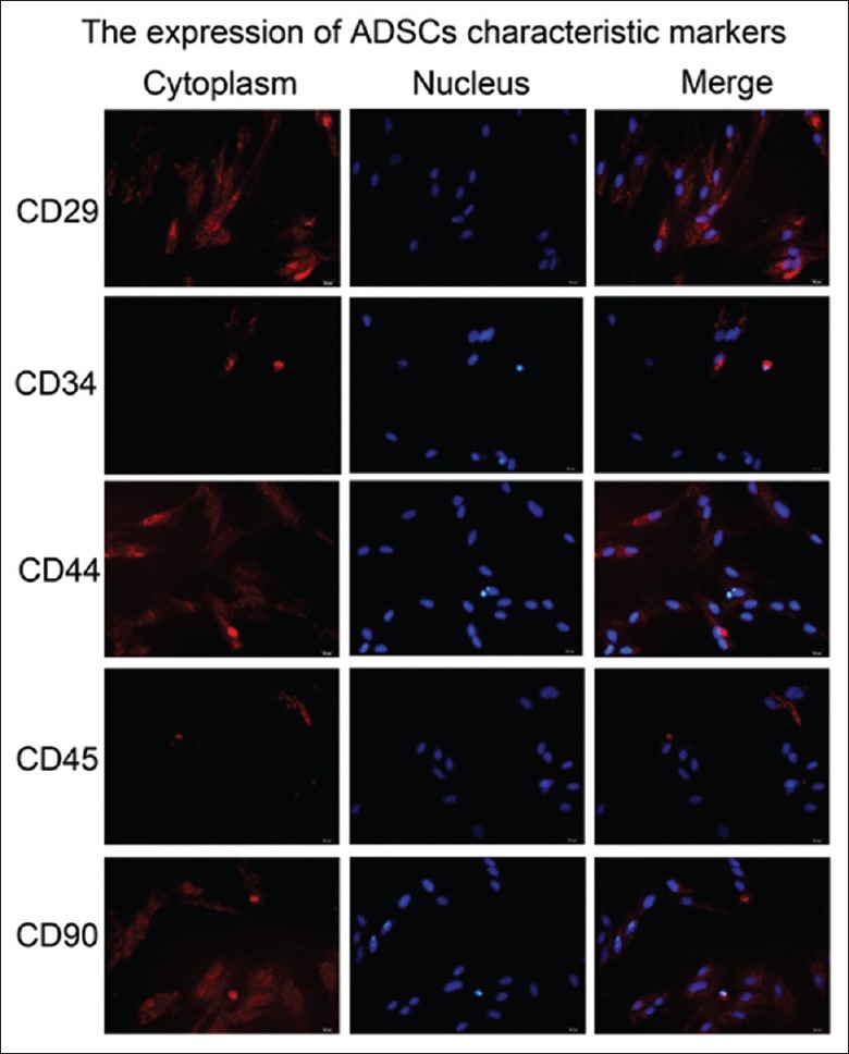
Figure 1.

The culture and identification of adipose-derived stem cells in vitro. The ADSCs morphology was observed under the microscope, the cells were spindle shaped and growing vigorously, and the mitotic figures were visible. The expression of the markers (CD29, CD90, CD34, and CD45) for ADSCs was shown as red fluorescence within the cells using IF analyses. The nuclei of the cells were stained blue with DAPI. Original magnification, ×400. ADSCs: Adipose-derived stem cells; IF: Immunofluorescence; DAPI: 4–6 diamidino-2-phenylinole.
