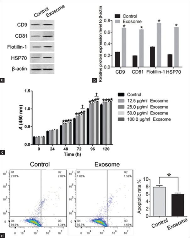
Figure 6.
Identification of exosomes with Western blotting and proliferation and apoptosis analyses of corneal stromal cells treated with exosomes in vitro using CCK-8 and annexin V-FITC methods. (a and b) Western blotting was used to analyze CD9, CD81, flotillin-1, and HSP70 in exosome. ADSCs alone were used as the control. The exosome-treated CSCs were defined as the exosome group. *P < 0.01 versus control. (c) CCK-8 Detection Kit was used to detect the proliferation of CSCs with exosomes. *P < 0.05 versus control. †P < 0.05 versus 50 μg/ml exosomes. (d) Annexin V-FITC Apoptosis Detection Kit was used to detect the apoptotic cells under control and exosome-treated conditions. All the bar graphs show the mean ± standard deviation in independent transfection experiments. CSCs alone were used as the control group, and the exosome-treated CSCs were defined as the exosome group. *P < 0.05 versus control. ADSCs: Adipose-derived stem cells; CSCs: Corneal stromal cells; CCK-8: Cell Counting Kit-8; FITC: Fluorescein isothiocyanate; PI: Propidium iodide.

